
Cadmium Accumulation in Some Organs of Rana Ridibunda Ridibunda Affect Erythrocytic Nuclear Abnormalities
DOI:
https://doi.org/10.26438/ijsrbs.v12i2.675Keywords:
Ranaridibunda, erythrocytic abnormalities, Genotoxicity, CadmiumAbstract
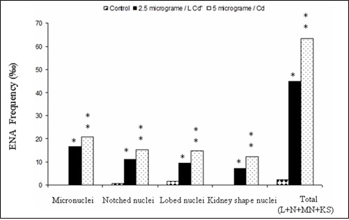
n the current research, accumulation of cadmium was investigated in various organs kidney, liver and the testis of Ranaridibunda exposed to diverse experimental levels of cadmium and erythrocytic abnormalities. Rana ridibunda species inhabits small shallow streams located in North Mosul / Iraq. Although cadmium level was not detectable in source water and expected to be with quality water guideline levels to protect the life in fresh freshwater proposed by the Agency of environmental protection (EPA), therefore the noticeable Cd level detected in experimental animals could pose danger to aquatic organisms. The Cd concentration in the different frog tissues was variable, the highest concentration was found in testis and the lowest in the liver. Frogs from treated groups had significantly higher hepatic (1.3232 2.1800, 3.5130 μg / g), renal (3.4556, 4.2850, 4.9992 μg /g) and testicular (3.5812, 4.8170, 5.5556 μg / g ) for 2.5, 5 and 10 Cd / µg /L respectively than those from the control group (1.0718, 1.9678, 3.2290 μg / g). There was erythrocytic nuclear abnormalities which significantly higher in number of Micronuclei (MN), notched nuclei (N), Lobed nuclei (L) and nuclei with kidney shaped (K) respectively were noted in the groups for 2.5 and 5 Cd / µg /L treated groups than that from natural sites, also with higher frequency significantly of immature erythrocyte with deleterious effect and totally erythrocytes demolished at 10 Cd / µg /g. This research found that cadmium accumulation of the different treatments frogs have higher in testicular, renal, hepatic and found a higher number of erythrocytic nuclear abnormalities when compared to frogs from natural habitat. Therefore, the accumulation of cadmium in the tissues that are used with concentrations and sampling periods is taken into consideration.
References
M.M.T. Jaber, Z.M. Al-Jumaa, S.K. Al-Taee, H.H. Nahi, M.O. Al-Hamdany, M.A. Al-Salh, B. Al-Mayahi, “Bioaccumulation of heavy metals and histopathological changes in muscles of common carp (Cyprinus carpio L.) in the Iraqi rivers,” Iraqi Journal of Veterinary Sciences, Vol.35, Issue.2, pp.245-249, 2021. http://dx.doi.org/10.33899/ijvs.2020.126748.1368
R.W. McDiarmid, J.C. Mitchell, “Diversity and distribution of amphibians and reptiles,” Ecotoxicology of Amphibians and Reptiles. SETAC Technical Publication Series, pp.15-69, 2000. https://pubs.er.usgs.gov/publication/5211075
M. Selvi, A. Gül, M. Yılmaz, “Investigation of acute toxicity of cadmium chloride (CdCl2· H2O) metal salt and behavioral changes it causes on water frog (Rana ridibunda Pallas, 1771),” Chemosphere, Vol.52, Issue.1, pp.259-263, 2003. http://dx.doi.org/10.1016/S0045-6535(03)00262-5
S.E. Mitchell, C.A. Caldwell, G. Gonzales, W.R. Gould, R. Arimoto, “Effects of depleted uranium on survival, growth, and metamorphosis in the African clawed frog (Xenopus laevis),” Journal of Toxicology and Environmental Health, Part A, Vol.68, Issue.11-12, pp.951-965, 2005. https://doi.org/10.1080/15287390590912595
V.K. Llewelyn, L. Berger, B.D. Glass, “Permeability of frog skin to chemicals: effect of penetration enhancers,” Heliyon, Vol.5, Issue.8, pp.1-12, 2019. http://dx.doi.org/10.1016/j.heliyon.2019.e02127
S.N. Stuart, J.S. Chanson, N.A. Cox, B.E. Young, A.S. Rodrigues, D.L. Fischman, R.W. Waller, “Status and trends of amphibian declines and extinctions worldwide.” Science, Vol.306, Issue.5702, pp.1783-1786, 2004. http://dx.doi.org/10.1126/science.1103538.
A. Zaghloul, M. Saber, S. Gadow, F. Awad, “Biological indicators for pollution detection in terrestrial and aquatic ecosystems,” Bulletin of the National Research Centre, Vol.44, Issue.127, pp.1-11, 2020. http://dx.doi.org/10.1186/s42269-020-00385-x
W. Adlassnig, S. Sassmann, A. Grawunder, M. Puschenreiter, A. Horvath, M. Koller-Peroutka, “Amphibians in metal-contaminated habitats,” Salamandra, Vol.49, Issue.3, pp.149-158, 2013. https://biorem.univie.ac.at
P. Thanomsangad, B. Tengjaroenkul, M. Sriuttha, L. Neeratanaphan, “Heavy metal accumulation in frogs surrounding an e-waste dump site and human health risk assessment,” Human and Ecological Risk Assessment: An International Journal, Vol.26, Issue.5, pp.1-16, 2019. http://dx.doi.org/10.1080/10807039.2019.1575181
A.A. Al-Kshab, O.Q. Fathi, “Determination of the lethal concentration 50%(LC50) of lead chloride and its accumulation in different organs of Gambusia affinis fish,” Iraqi Journal of Veterinary Sciences, Vol.2, pp.361-367, 2021. http://dx.doi.org/10.33899/ijvs.2020.126853.1401
L. Bervoets, R. Blust, “Metal concentrations in water, sediment and gudgeon (Gobio gobio) from a pollution gradient: relationship with fish condition factor,” Environmental pollution, Vol.126, Issue.1, pp.9-19, 2003. http://dx.doi.org/10.1016/s02697491
J. Burger, J. Snodgrass, “Metal levels in southern leopard frogs from the Savannah River Site: location and body compartment effects,” Environmental research, Vol.86, Issue.2, pp.157-166, 2001. http://dx.doi.org/10.1006/enrs.2001.4245
N.S. Loumbourdis, A.K. Vogiatzis, “Impact of cadmium on liver pigmentary system of the frog Ranaridibunda,” Ecotoxicol Environ Saf, Vol.53, Issue,1, pp.52–58, 2002. http://dx.doi.org/10.1007/s11356-016-6153-z.
R. Jaafar, “Bioremediation of lead and cadmium and the strive role of Pediococcus pentosaceus probiotic,” Iraqi Journal of Veterinary Sciences, Vol.34, Issue.1, pp.51-57, 2020. http://dx.doi.org/10.33899/ijvs.2019.125581.1092.
I. Bochenek, M. Protasowicki, E. Brucka-Jastrzębska, “Studies on the bioavailability of heavy metals (Cd, Pb, Cu, Zn) from bottom sediments to guppies,” Poecilia reticulata Peters. Fisheries & Aquatic Life, Vol.16, Issue.2, pp.155-166, 2008. http://dx.doi.org/10.2478/s10086-008-0013-5
G. Genchi, M.S. Sinicropi, G. Lauria, A. Carocci, A. Catalano, “The effects of cadmium toxicity,” International journal of environmental research and public health, Vol.17, Issue.11, p.3782, 2020. http://dx.doi.org/10.3390/ijerph17113782.
B. Yeşilbudak, C. Erdem, “Cadmium accumulation in gill, liver, kidney and muscle tissues of common carp, Cyprinus carpio, and Nile tilapia, Oreochromis niloticus,” Bulletin of environmental contamination and toxicology, Vol.92, pp.546-550, 2014. http://dx.doi.org/10.1007/s00128-014-1228-3.
S.A. Mustafa, “Histopathology and heavy metal bioaccumulation in some tissues of Luciobarbusxanthopterus collected from Tigris River of Baghdad, Iraq,” The Egyptian Journal of Aquatic Research, Vol.46, Issue.2, pp.123-129, 2020 . http://dx.doi.org/10.1016/j.ejar.2020.01.004.
H. Zhang, C. Cai, C. Shi, H. Cao, Z. Han, X. Jia, “Cadmium-induced oxidative stress and apoptosis in the testes of frog Rana limnocharis,” Aquatic toxicology, Vol.122, pp.67-74, 2012. http://dx.doi.org/10.1016/j.aquatox.2012.05.014
F.H. Pough, “Amphibian biology and husbandry. ILAR journal / National Research Council,” Institute of Laboratory Animal Resources, Vol.48, Issue.3, pp.203-13, 2007. http://dx.doi.org/10.1093/ilar.48.3.203.
B. Jezierska, M. Witeska, “The metal uptake and accumulation in fish living in polluted waters,” In Soil and water pollution monitoring, protection and remediation, Springer Netherlands, pp.107-114, 2006. http://dx.doi.org/10.1007/978-1-4020-4728-2_6 .
X. Jia, H. Zhang, X. Liu, “Low levels of cadmium exposure induce DNA damage and oxidative stress in the liver of Oujiang colored common carp Cyprinus carpio var. color.,” Fish physiology and biochemistry, Vol.37, pp.97-103, 2011. http://dx.doi.org/10.1007/s10695-010-9416-5
R. Martí-Cid, G. Perelló, J.L. Domingo, “Dietary exposure to metals by individuals living near a hazardous waste incinerator in Catalonia, Spain: temporal trend,” Biological Trace Element Research, Vol.131, pp.245-254, 2009. http://dx.doi.org/10.1007/s12011-009-8368-z.
K.R. Carrasco, K.L. Tilbury, M.S. Myers, “Assessment of the piscine micronuclei test as an in situ biological indicator of chemical contaminant effects,” Canadian Journal of Fisheries and Aquatic Sciences, Vol.47, Issue.11, pp.2123-2136, 1990. https://doi.org/10.1139/f90-237
S.M. Marques, S.C. Antunes, H. Pissarra, M.L. Pereira, F. Gonçalves, R. Pereira, “Histopathological changes and erythrocytic nuclear abnormalities in Iberian green frogs (Rana perezi Seoane) from a uranium mine pond,” Aquatic Toxicology, Vol.91, Issue.2, pp.187-195, 2009. http://dx.doi.org/10.1016/j.aquatox.2008.04.010.
O. Faroon, A. Ashizawa, S. Wright, P. Tucker, K. Jenkins, L. Ingerman, C. Rudisill, “Toxicological profile for cadmium,” Atlanta (GA), 2013.
P. B. Tchounwou, C.G. Yedjou, A.K. Patlolla, D.J. Sutton, “Heavy Metals Toxicity and the Environment,” Environmental toxicology, Vol.101, pp.133–164, 2012. http://dx.doi.org/10.1007/978-3-7643-8340-4_6.
A.R. Ahmed, “Evaluation of the heavy metal content in the muscle tissue of common carp (Cyprinus carpio L.) reared in groundwater in Basrah province, Iraq,” Iraqi Journal of Veterinary Sciences, Vol.35, Issue.1, pp.157-161, 2021. http://dx.doi.org/10.33899/ijvs.2020.126491.1336.
N.U. Rehman, M.N. Ansari, M.A. Ganaie, H.A. Madkhali, A.S. Saeedan, F. Imam, A.M. Hamad, “Cadmium-induced Hepatotoxicity and Oxidative Stress in Rats: Protection by Roflumilast via NF-kappa B and HO-1 Pathway,” International Journal of Pharmacology, Vol.16, Issue.2, pp.154-163, 2020. http://dx.doi.org/10.3923/ijp.2020.154.163.
A.L.R. Mercé, "Contaminated Waters and Depleted Soils: Impact in Nutrition II Biochemical Detoxification Pathways". EC Nutrition, 19, 1–29. 2024 https://doi.org/10.31031/ACAM.2023.07.000678
S. Satarug, "Is Chronic Kidney Disease due to Cadmium Exposure Inevitable and Can it be Reversed", Biomedicines, 12(4), 718, 2024; https://doi.org/10.3390/biomedicines12040718
M. Nasiadek, J. Stragierowicz, A. Kilanowicz, "An Assessment of Metallothionein–Cadmium Binding in Rat Uterus after Subchronic Exposure Using a Long–Term Observation Model". International Journal of Molecular Sciences, 23(23), 15154. 2022. https://doi.org/10.3390/ijms232315154
C.J. Bautista, N. Arango, P. Consuelo, I.B. Mitre-Aguilar, J. Trujillo, V. Ramírez, "Mechanism of Cadmium-induced nephrotoxicity". Toxicology, 502 153726. 2024 https://doi.org/10.1016/j.tox.2024.153726
N.S. Loumbourdis, D. Wray, “Heavy-metal concentration in the frog Rana ridibunda from a small river of Macedonia, Northern Greece,” Environment international, Vol.24, Issue.4, pp.427-431, 1998. http://dx.doi.org/10.1016/S0160-4120
C.S. Pérez-Coll, J. Herkovits, O. Fridman, P. Daniel, J.L. D'eramo, “Metallothioneins and cadmium uptake by the liver in Bufo arenarum,” Environmental Pollution, Vol.97, Issue.3, pp.311-315, 1997. http://dx.doi.org/10.1289/ehp.6647.
C.M. Foran, B.N. Peterson, W.H. Benson, “Influence of parental and developmental cadmium exposure on endocrine and reproductive function in Japanese medaka (Oryzias latipes),” Comparative Biochemistry and Physiology Part C: Toxicology & Pharmacology, Vol.133, Issue.3, pp.345-354, 2002. http://dx.doi.org/10.1293/tox.2017-0015.
M. Chandel, G.C. Jain, “Toxic effects of transition metals on male reproductive system: A review,” Journal of Environmental and Occupational Science, Vol.3, Issue.4, pp.1-11, 2014. http://dx.doi.org/10.5455/jeos.20140929042630
M. Fenech, “The in vitro micronucleus technique,” Mutation Research/Fundamental and Molecular Mechanisms of Mutagenesis, Vol.455, No.1-2, pp.81-95, 2000. https://doi.org/10.1016/S0027-5107.
T. Stoiber, D. Bonacker, K.J. Böhm, H.M. Bolt, R. Thier, G.H. Degen, E. Unger, “Disturbed microtubule function and induction of micronuclei by chelate complexes of mercury (II),” Mutation Research/Genetic Toxicology and Environmental Mutagenesis, Vol.563, Issue.2, pp.97-106, 2004. http://dx.doi.org/10.1016/j.mrgentox.2004.06.009
F. Ayllon, E. Garcia-Vazquez, “Induction of micronuclei and other nuclear abnormalities in European minnow Phoxinus phoxinus and mollie Poecilia latipinna: an assessment of the fish micronucleus test,” Mutation Research/Genetic Toxicology and Environmental Mutagenesis, Vol.467, Issue.2, pp.177-186, 2000. http://dx.doi.org/10.1016/s1383-5718
L. Serrano‐García, R. Montero‐Montoya, “Micronuclei and chromatid buds are the result of related genotoxic events,” Environmental and molecular mutagenesis, Vol.38, Issue.1, pp.38-45, 2001. http://dx.doi.org/10.1002/em.1048
S. Guilherme, M. Válega, M.E. Pereira, M.A. Santos, M. Pacheco, “Erythrocytic nuclear abnormalities in wild and caged fish (Liza aurata) along an environmental mercury contamination gradient,” Ecotoxicology and Environmental Safety, Vol.70, Issue.3, pp.411-421, 2008. http://dx.doi.org/10.1016/j.ecoenv.2007.08.016
R.H. Foote, E.d. Carney, “The rabbit as a model for reproductive and developmental toxicity studies,” Reproductive Toxicology, Vol.14, Issue.6, pp.477-93, 2000. http://dx.doi.org/10.1016/s0890-6238.
H. Wang, P. Huang, T. Lie, F. Shi, “Reproductive toxicity of acrylamide-treated male rats,” Reproductive Toxicology, Vol.29, Issue.2, pp.225-30, 2009. http://dx.doi.org/10.1016/j.reprotox.2009.11.002.
D.R. Livingstone, P.G. Martinez, X. Michel, J.F. Narbonne, S. O'hara, D. Ribera, G.W. Winston, “Oxyradical production as a pollution-mediated mechanism of toxicity in the common mussel, Mytilus edulis L., and other molluscs,” Functional ecology, pp.415-424, 1990. https://doi.org/10.2307/2389604.
S.J. Stohs, D. Bagchi, E., Hassoun, M. Bagchi, “Oxidative mechanisms in the toxicity of chromium and cadmium ions,” Journal of environmental pathology, toxicology and oncology, Vol.20, Issue.2, 2001. http://dx.doi.org/10.1615/JEnvironPatholToxicolOncol.v20.i2.10
Downloads
Published
How to Cite
Issue
Section
License
Copyright (c) 2025 Semaa Baker, Ammar Alhaaik , Hussein Mohammed-Ali , Talib Ali

This work is licensed under a Creative Commons Attribution 4.0 International License.
Authors contributing to this journal agree to publish their articles under the Creative Commons Attribution 4.0 International License, allowing third parties to share their work (copy, distribute, transmit) and to adapt it, under the condition that the authors are given credit and that in the event of reuse or distribution, the terms of this license are made clear.